Education
Minimally Invasive Decompression for Spinal Stenosis
H. Louis Harkey, MD, Professor of Neurosurgery
University of Mississippi Medical Center, Jackson
Position – "Knee-chest prone." The patient is positioned on the Andrews Table with the hips and knees flexed 90º and the lumbar spine parallel to the floor. In this position, the weight of the patient is distributed to the sternum and knees, leaving the abdomen completely free.
 Localization - C-arm fluoroscopy (or lateral x-ray) imaging precisely localizes the entry point, which is determined by the level of compression on clinical exam and preoperative imaging. The goal is to center the retractor over the disc space if compression is limited to a single level or over the intervening lamina if there are two adjacent levels of compression.
Incision – A 1-inch incision is made in the skin ½ inch lateral to the midline. The side of exposure is determined by the most symptomatic side or the side of greatest compression on diagnostic imaging. The exposure extended to the lumbar fascia.
Dissection - The fascia is opened linearly with a 1-inch incision; taking care not to cut the underlying muscle. Using blunt dissection with a small flat instrument (e.g. freer) the muscle is separated form the medial facial flap to the midline. This plane can be followed within the muscle fascia all the way to the lamina. This is intrafascial dissection, not subperiosteal. On occasion, muscle fibers must be sharply dissected from their medial attachments, but for the most part, a distinct plane can be followed with blunt dissection.
A speculum retractor (Aesculap Caspar MLD) is inserted into the opening created by the initial dissection. A lateral counter retractor is applied after opening the speculum. The final position should be confirmed with an intraoperative X-ray.
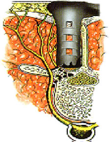 From this point the microscope is used for visualization. Any remaining soft tissue can be removed with electrocautery. The exposure includes the base of the spinous processes, lamina and facet joint. Additional rostral and caudal exposure can be achieved by changing the angle of the speculum.
A high speed drill and Kerrison rongeurs are used to create the laminotomy, foramenotomy and facetectomy (if necessary).
Angling the speculum up and down allows the surgeon to decompress both the exiting and traversing nerve roots (two intervals). Angling the speculum medially and undercutting the lamina/spinous process allows the surgeon to achieve a central decompression and even decompress the nerve roots on the opposite side.
Closure – The fascia and subcutaneous layers are closed with absorbable suture. The skin is sealed with tissue glue, which serves the dual purpose of skin approximation and wound dressing.